

Mehr zum Buch
Since the small bowel except the duodenum and (1961), Pygott et al. (1960), Gianturco (1967) terminal ileum is largely inaccessible during en and Bilbao et al. (1967). doscopic examination, radiology of the small Sellink, however, was really responsible for bowel attains special significance as a diagnostic the widespread recognition of enteroclysis method. Owing to the length and position of (1971, 1974, 1976). In spite of the increasing this organ, good images are difficult to obtain. popularity of this method, the necessity for sub Furthermore, the considerable variation oftran stituting this apparently viable method for the sit time, unpredictable response of the contrast peroral examination is still equivocal (Rabe medium, and superimposition with the filled etal. 1981; Fried etal. 1981; Maglinte etal. loops make small bowel radiology difficult. As 1982; Ott et al. 1985). Comparisons of both methods, however, (Fleckenstein and Pedersen a result, few radiologists specialize in this field. With the exception of Crohn's disease, disorders 1975; Sanders and Ho 1976; Ekberg 1977; Val lance 1980) have confirmed the superiority of of the small bowel are relatively rare. Thus, not many clinicians and radiologists are interested enteroclysis. It achieves a high accuracy (Antes in the small intestine. and Lissner 1983)."
Buchkauf
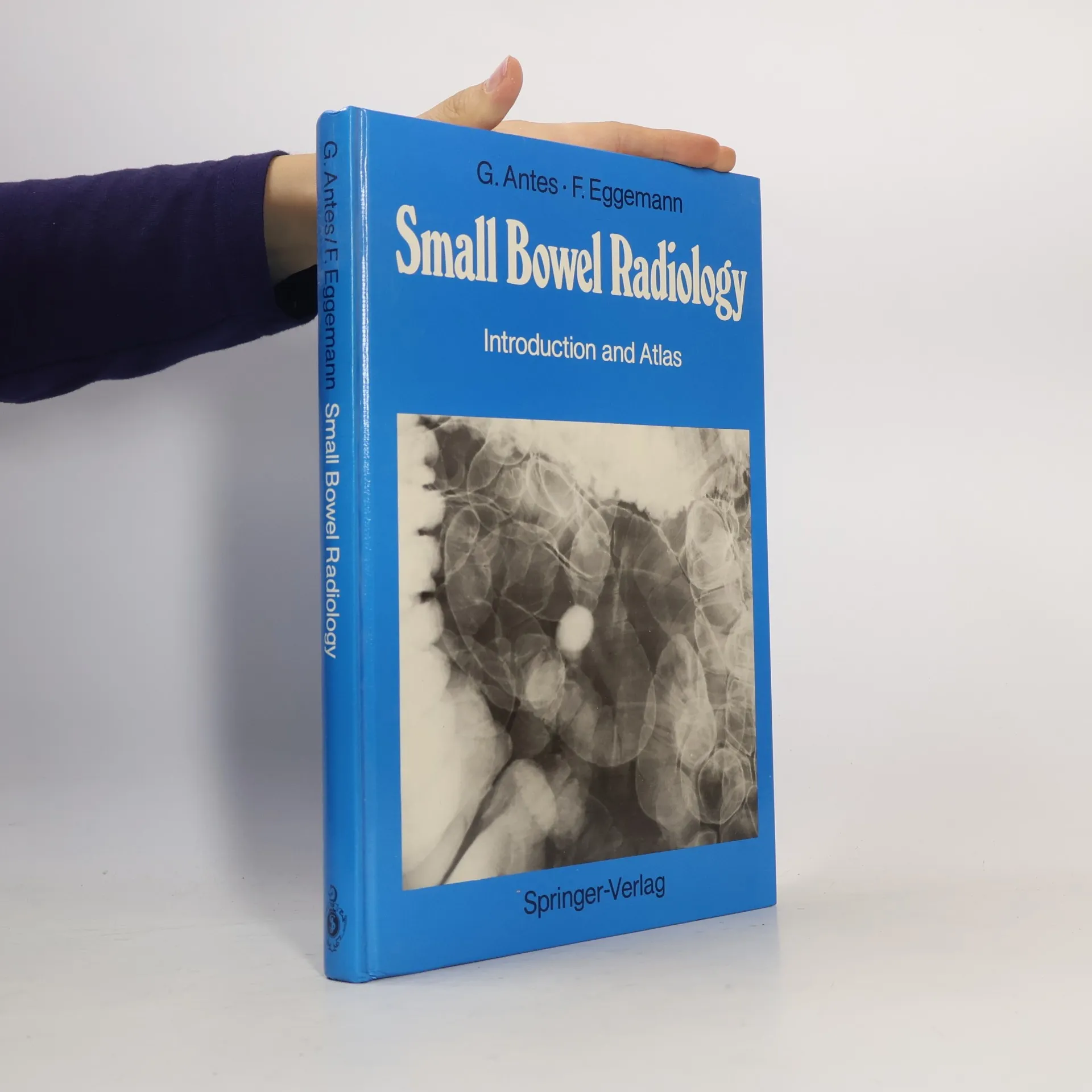
Small Bowel Radiology, Günther Antesberger, Josef Lissner, Franz Eggemann
- Sprache

- Erscheinungsdatum
- 1988
- Einband
- (Hardcover),
- Buchzustand
- Gebraucht - Gut
- Preis
- 25,49 €inkl. MwSt.
Keiner hat bisher bewertet.
- Titel
- Small Bowel Radiology
- Untertitel
- Introduction and Atlas
- Sprache
- Englisch
- Autor*innen
- Günther Antesberger, Josef Lissner, Franz Eggemann
- Verlag
- Springer
- Erscheinungsdatum
- 1988
- Einband
- Hardcover
- Seitenzahl
- 207
- ISBN10
- 3540152636
- ISBN13
- 9783540152637
- Reihe
- Schlagwörter
- Medizin, Krebs, Tumoren, Chirurgie, Biochemie, Radiologie, Gastroenterologie
- Beschreibung
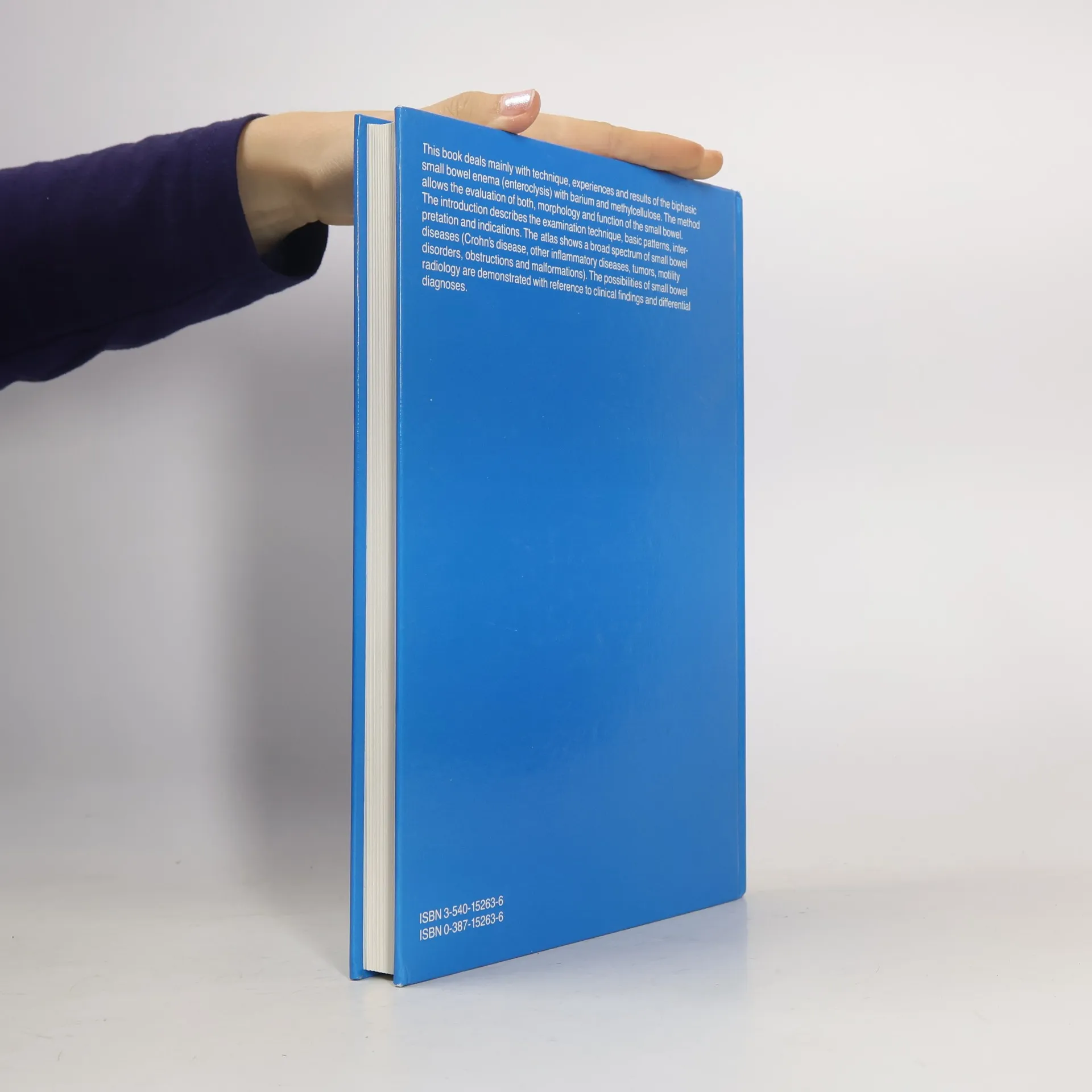
- Since the small bowel except the duodenum and (1961), Pygott et al. (1960), Gianturco (1967) terminal ileum is largely inaccessible during en and Bilbao et al. (1967). doscopic examination, radiology of the small Sellink, however, was really responsible for bowel attains special significance as a diagnostic the widespread recognition of enteroclysis method. Owing to the length and position of (1971, 1974, 1976). In spite of the increasing this organ, good images are difficult to obtain. popularity of this method, the necessity for sub Furthermore, the considerable variation oftran stituting this apparently viable method for the sit time, unpredictable response of the contrast peroral examination is still equivocal (Rabe medium, and superimposition with the filled etal. 1981; Fried etal. 1981; Maglinte etal. loops make small bowel radiology difficult. As 1982; Ott et al. 1985). Comparisons of both methods, however, (Fleckenstein and Pedersen a result, few radiologists specialize in this field. With the exception of Crohn's disease, disorders 1975; Sanders and Ho 1976; Ekberg 1977; Val lance 1980) have confirmed the superiority of of the small bowel are relatively rare. Thus, not many clinicians and radiologists are interested enteroclysis. It achieves a high accuracy (Antes in the small intestine. and Lissner 1983)."