Netter Grundlagen der Wissenschaft Reihe
Diese Serie vermittelt ein grundlegendes Verständnis wesentlicher wissenschaftlicher Konzepte durch klare, zugängliche Erklärungen und anschauliche Illustrationen. Komplexe biologische und physiologische Systeme werden sorgfältig zerlegt und leicht verständlich gemacht. Diese Ressource ist von unschätzbarem Wert für Studierende der Medizin und Gesundheitswissenschaften sowie für alle, die sich für die Komplexität des menschlichen Körpers interessieren.

Empfohlene Lesereihenfolge
El Atlas Práctico de Anatomía Ortopédica, dirigido por Jon C. Thompson, es una obra esencial para estudiantes de Traumatología y Ortopedia. Esta segunda edición incluye más imágenes anatómicas y amplía secciones sobre procedimientos quirúrgicos. Las ilustraciones de Netter y el diseño de tablas facilitan la revisión de patologías ortopédicas.
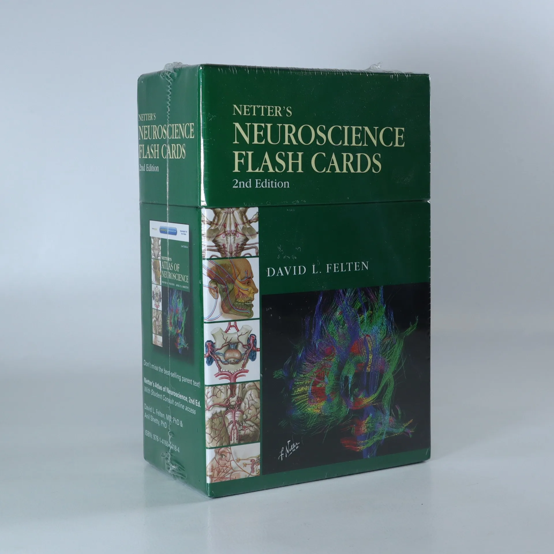
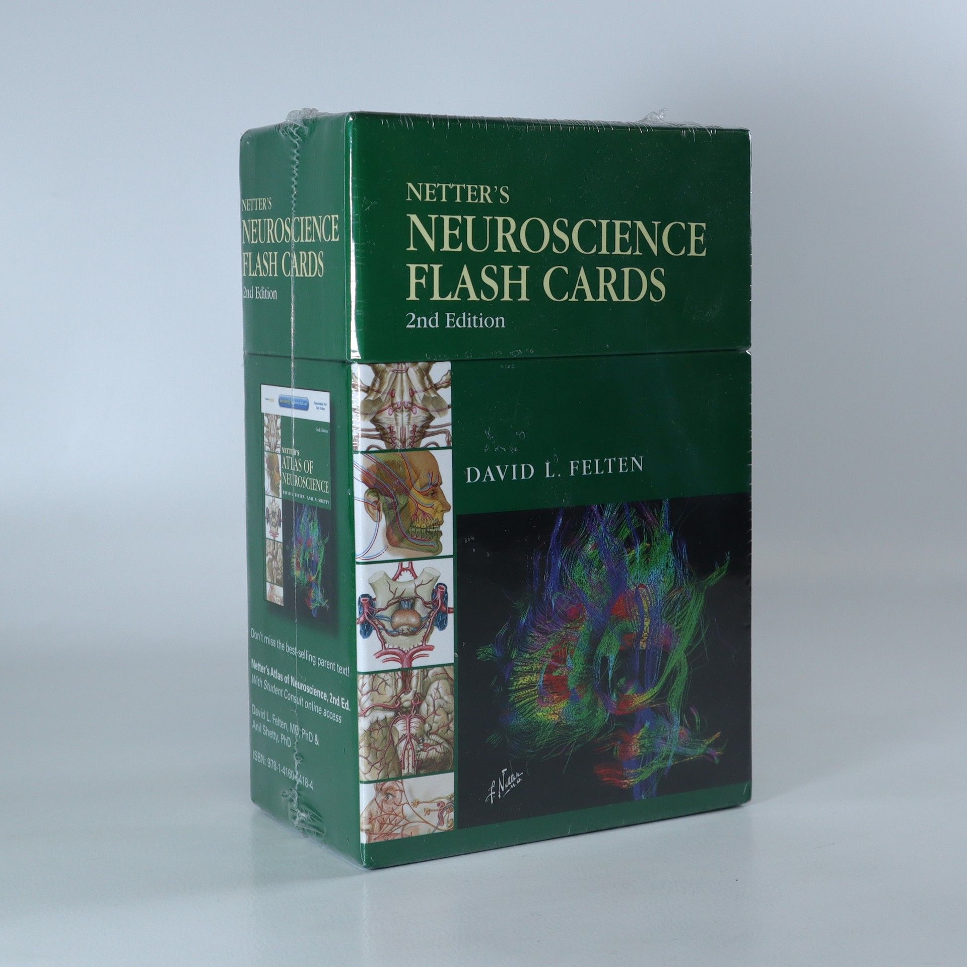
Netter's neuroscience flash cards
- 450 Seiten
- 16 Lesestunden
450 pages Master the structures and clinical points most important to a basic medical neuroscience course. Use the cards before exams or boards, or throughout clinical rotations, residency, or in practice for a fast review of the nervous system. Cross-reference with Netter's Atlas of Neuroscience, 2nd Ed. for further information on any topic. Review clinical 'pearls' and helpful summary information on the back of each card to understand the clinical implications of neuroscience concepts. Use the card set's pre-punched holes and convenient binding ring to carry selected groups of flash cards with you anywhere. View more neuroimaging examples to assess your grasp of this important subject. Make clinically important correlations in neuroanatomy, cell biology, and neurophysiology.
Netter's Advanced Head and Neck Flash Cards
- 540 Seiten
- 19 Lesestunden
Recto of each card is a colored illustration with salient anatomical features numbered; verso lists the names of the numbered features and gives important facts about their formation and about associated features. One or more cards in sections 1 - 10 illustrate and discuss clinical correlates.
W Atlasie anatomii radiologicznej Nettera zestawiono obrazy uzyskane w badaniach radiologicznych - takich jak rezonans magnetyczny, badanie ultrasonograficzne i tomografia komputerowa - ze znakomitymi pracami mistrza ilustracji medycznych, dra Franka H. Nettera. Omówiono też kliniczne znaczenie przedstawianych struktur.
Netter's Atlas of Human Embryology
- 267 Seiten
- 10 Lesestunden
Here's a rich pictorial review of normal and abnormal human prenatal development. For each body system or region, you'll find a brief description of the developmental plan, with key concepts and terminology, followed by discussions of histological principles, the classification of congenital defects, and basic cellular, molecular, and genetic concepts.An emphasis on morphological patterns in the embryo and fetus makes it easy to understand the structure and function of the adult body and the embryonic basis of birth defects.Summary tables and terminology sections at the end of each chapter, plus an appendix with all major congenital defects and their embryonic basis, make it easy to review course material and prepare for the USMLE.

Netter's Head and Neck Anatomy for Dentistry
- 712 Seiten
- 25 Lesestunden
This guide offers a clear and visual approach to understanding anatomy specifically tailored for dental professionals. It emphasizes clinically relevant details that are essential for effective practice in dentistry. The book combines illustrations and concise explanations to enhance learning and retention, making it an invaluable resource for students and practitioners alike.
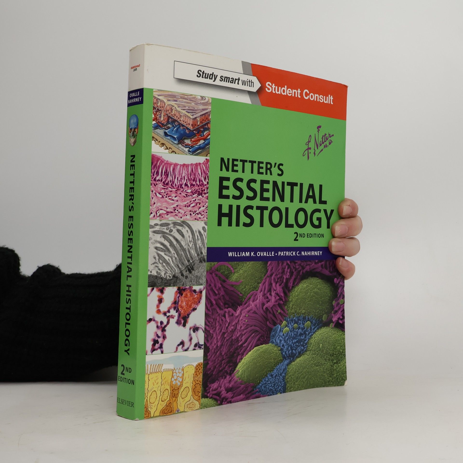
Netter's Essential Histology
- 536 Seiten
- 19 Lesestunden
Netter's Essential Histology integrates gross anatomy and embryology with classic histology slides and cutting-edge scanning electron microscopy to give you a rich visual understanding of this complex subject. This histology textbook-atlas has a strong anatomy foundation and utilizes a variety of visual elements - including Netter illustrations and light and electron micrographs - to teach you the most indispensable histologic concepts and their clinical relevance. Excellent as both a reference and a review, Netter's Essential Histology will serve you well at any stage of your healthcare career. Gain a rich understanding of this vital subject through the succinct explanatory histology text. Learn to recognize both normal and diseased structures at the microscopic level with the aid of succinct explanatory text as well as numerous clinical boxes. Access the entire contents and ancillary components online at Student Consult, view images and histology slides at different magnifications, and watch new narrated video overviews of each chapter. Take your learning one step further with the purchase of Netter's Histology Flash Cards (sold separately), designed to reinforce your understanding of how the human body works in health as well as illness and injury. Thoroughly comprehend how function is linked to structure through brand-new electron micrographs, many of which have been enhanced and colorized to show ultra-structures in 3D.
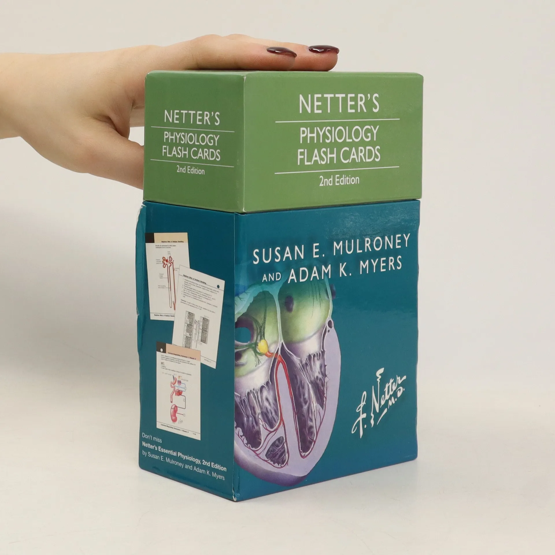
Netter's Physiology Flash Cards
- 450 Seiten
- 16 Lesestunden
Find out why more students prefer Netter's Physiology Flash Cards, 2nd Edition, for a quick review and self-test of human physiology essentials. These bestselling, beautifully illustrated cards are fully up to date, with images and questions on the front, answers and clinical correlations on the reverse. Over 200 hole-punched cards, organized by body system, provide concise, quick-access information on key physiology concepts for the perfect, portable review. High-quality Netter and Netter-style illustrations enhance learning. More than a dozen new cards offer expanded coverage of blood and lymph nodes while more clinical correlates throughout help you apply what you've learned. Cross-referenced to Netter's Essential Physiology, 2nd Edition, but also highly effective when used with any preferred physiology text. Ideally suited for individual or group study - and universally appreciated by undergraduate, nursing, allied health, and medical students! Over a dozen new cards - including expanded coverage of blood and lymph and more clinical correlates Cross-references to the 2e of the parent book Electronic options (**not free with print**)

Atlas of Human Anatomy uses Frank H. Netter, MD's detailed illustrations to demystify this often intimidating subject, providing a coherent, lasting visual vocabulary for understanding anatomy and how it applies to medicine. This 5th Edition features a stronger clinical focus-with new diagnostic imaging examples-making it easier to correlate anatomy with practice. Student Consult online access includes supplementary learning resources, from additional illustrations to an anatomy dissection guide and more. Netter. It's how you know.
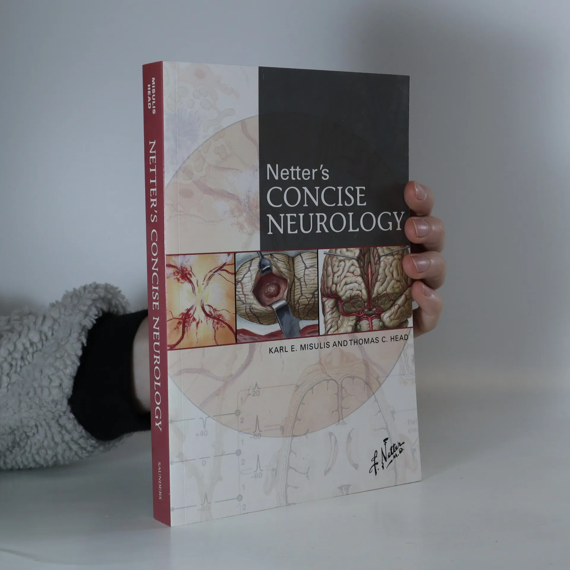
Netter's concise neurology
- 586 Seiten
- 21 Lesestunden
More than 200 exquisite, hand-painted illustrations - created by, and in the style of, master medical illustrator Frank H. Netter, MD - capture the essential clinical aspects of over 200 major neurologic disorders seen in hospital and office practice. A masterful combination of artwork, succinct text, and tables, together with a highly compact format, deliver quick and convenient access to vital clinical knowledge!
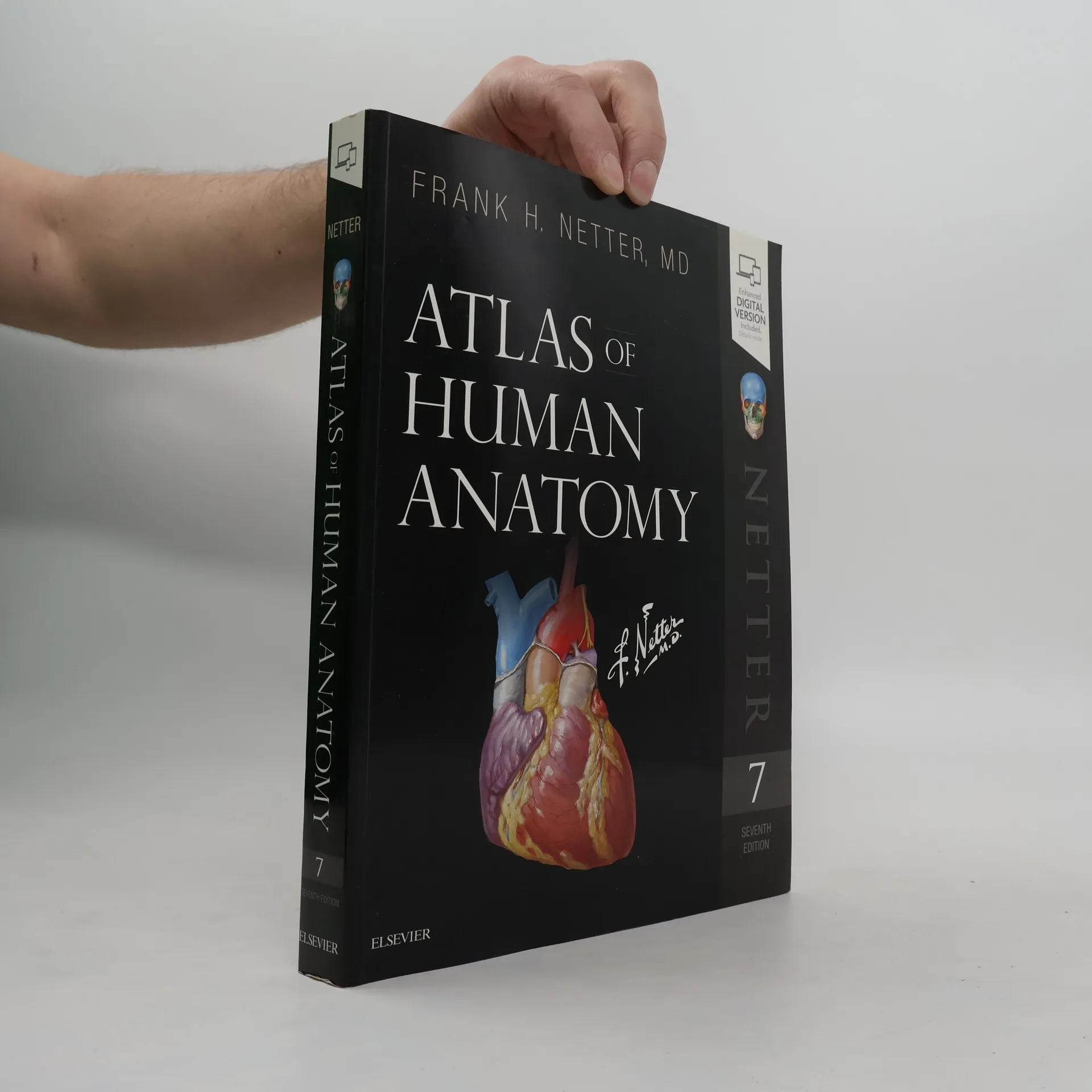
Atlas of Human Anatomy 7
- 672 Seiten
- 24 Lesestunden
"The only anatomy atlas illustrated by physicians, Atlas of Human Anatomy, 7th edition, brings you world-renowned, exquisitely clear views of the human body with a clinical perspective. In addition to the famous work of Dr. Frank Netter, you'll also find nearly 100 paintings by Dr. Carlos A. G. Machado, one of today's foremost medical illustrators. Together, these two uniquely talented physician-artists highlight the most clinically relevant views of the human body. In addition, more than 50 carefully selected radiologic images help bridge illustrated anatomy to living anatomy as seen in everyday practice"--Publisher's description

Netter's Head and Neck Anatomy for Dentistry
- 659 Seiten
- 24 Lesestunden
Netter's Head and Neck Anatomy for Dentistry, by Neil S. Norton, PhD, uses more than 600 full-color images from the Netter Collection to richly depict all of the key anatomy that's relevant to clinical practice. This new edition takes your knowledge further than ever with more Netter illustrations; addition of over 20 cone beam CT images; new chapters on the upper limbs, thorax, and abdomen; and more than 100 multiple-choice questions. Whether for your dental anatomy course, board review, or as a handy reference in your dental office, this concise, visual guide is an excellent anatomy atlas and quick reference for students and professionals in dentistry and dental hygiene. Identify clinically relevant anatomy with Netter illustrations highlighted and modified for dentistry. See the practical important of anatomy from illustrated clinical examples in each chapter. Review essential concepts easily with tables that display the maximum amount of information in an at-a-glance format. Master anatomy for the head and neck and beyond, including upper limbs, thorax, and abdomen. Stay current on hot topics like cone beam CT imaging, intraoral injections, and anesthesia. Recognize the context and clinical relevance of head and neck anatomy through additional coverage of dental procedures. Prepare effectively for the dental boards with over 100 multiple-choice questions.

This portable, full-color resource is essential for students and professionals during orthopaedic rotations and practice. Jon C. Thompson updates diagnostic and treatment algorithms while maintaining a user-friendly table format. Enhanced with additional artwork from the Netter Collection and new radiologic images, it effectively illustrates key clinical correlations and anatomical applications. It serves as a quick and memorable review of orthopaedic anatomy, making it a valuable tool for anyone in the field.